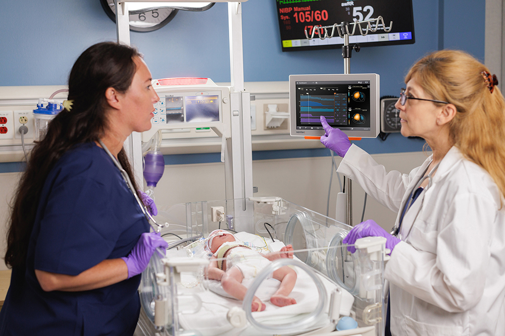

For individuals with sleep-disordered breathing — a group of conditions that cause abnormal breathing patterns during sleep — oxygenation, ventilation, and overall sleep quality can all be disrupted. These conditions are common and often underdiagnosed, yet their impact is significant; when left untreated, they’re associated with serious health consequences, including increased mortality.¹
In the sleep lab, carbon dioxide (CO2) monitoring can reveal breathing disturbances during overnight studies that might be missed when relying on oxygen (O2) levels alone. With this visibility into patients’ ventilation, sleep teams can make more informed decisions both during and after studies, ultimately supporting better outcomes for their patients.
Why Should CO2 Be Monitored During Sleep Studies?
When discussing disordered breathing, it helps to start with what normal breathing looks like. At its core, breathing has two essential components:
- Oxygenation: This is the process of getting oxygen into the bloodstream. In healthy patients, air reaches the alveoli, oxygen moves into the blood, and tissues throughout the body get what they need to produce energy, support organ function, and keep you alert and healthy.
- Ventilation: This is the movement of air in and out of the lungs. When ventilation is working properly, each breath brings in fresh oxygen and removes CO2, helping maintain a stable acid–base balance and prevent CO2 buildup in the body.
Both oxygenation and ventilation are essential, yet many sleep labs routinely measure only oxygenation (via pulse oximetry). When oxygen levels look stable, it’s easy to assume ventilation is adequate, but that’s not always the case.
Oxygenation does not reliably reflect ventilation, with several studies highlighting this gap:
- In patients with neuromuscular disorders, one study found that 41% of those with nocturnal alveolar hypoventilation had CO2 abnormalities, even though most had normal oxygen saturation readings.²
- In a study of patients on or recently weaned from CPAP, NIV, or nasal cannula, pulse oximetry detected only 2 of 19 gas-exchange abnormalities across 13 overnight recordings.³
These CO2 abnormalities are more common in sleep-lab populations than some may realize. Research shows that up to 46% of pediatric patients with sleep-disordered breathing experience a nighttime rise in transcutaneous CO2 greater than 10 mmHg,4 and that 17% of adults with obstructive sleep apnea may also have hypercapnia.5 And when monitoring just O2, these abnormalities could go unnoticed.
Overlooking CO2 in sleep lab patients can have serious consequences, as hypercapnia (elevated CO2), the result of inadequate ventilation, has been associated with:
- Cognitive function deficits6
- Increased exacerbations in patients with COPD7
- Higher risk of mortality8
Methods of CO2 Monitoring in the Sleep Lab
Monitoring CO2 plays an important role in overnight sleep studies, and there are several ways to do this in the sleep lab. Each method has its own benefits and limitations, which sleep teams should consider when selecting the most appropriate option for their practice.
Arterial Blood Gas (ABG)
Arterial blood gas tests provide a direct measurement of PaCO2 using a blood sample, typically taken from the radial artery at the wrist or the brachial artery at the inner elbow.
The pros:
- Industry standard for accuracy
- Provides multiple parameters in a single measurement (pH, CO2, O2, HCO3⁻, electrolytes)
- Direct assessment of arterial blood gas values
The limitations:
- Invasive and carries the risk of infection
- Only provides a point-in-time measurement
- Daytime readings do not reflect or predict nighttime events
- Requires skilled handling and timely processing to ensure accurate results
In summary…
While ABGs deliver the most accurate measurement of CO2, the procedure is invasive and only reflects the moment the sample is taken — potentially leaving hours of unmonitored time throughout the night. Not knowing what occurs between draws can have meaningful consequences for patients, as important fluctuations in CO2 may go undetected. Because repeated arterial sticks are both painful and impractical, ABGs are rarely used for monitoring during sleep studies, though some labs still rely on them in select situations.
End-Tidal CO2 Monitoring (Capnography)
End-tidal CO2 (EtCO2) monitoring, or capnography, is a continuous monitoring method that measures the level of carbon dioxide at the end of an exhaled breath, typically using a nasal cannula.
The pros:
- Noninvasive
- Ideal for diagnostic studies if there’s not a significant obstruction in airflow
- Delivers a breath-by-breath waveform
The limitations:
- Accuracy can be affected by leakage around uncuffed endotracheal tubes
- Can underestimate CO2 in patients with ventilation-perfusion (V/Q) mismatch (common in patients with apnea, obesity, and COPD)
- Incompatible with noninvasive ventilation (NIV) modalities
- Cannula may require occasional adjustment overnight due to patient movement and can cause irritation
- Cannula can become obstructed by secretions
In summary…
End-tidal CO2 monitoring can provide continuous insight into a patient’s ventilation, but several factors can limit its reliability and practicality in the sleep lab. Its use can be especially challenging during split-night studies, as EtCO2 often becomes unreliable when CPAP or other forms of noninvasive ventilation are applied, leaving part of the night without accurate data. These limitations should be considered when selecting a monitoring method or interpreting overnight study results, to ensure the patient’s ventilation is accurately assessed throughout the night.
Transcutaneous CO2 (tcPCO2) Monitoring
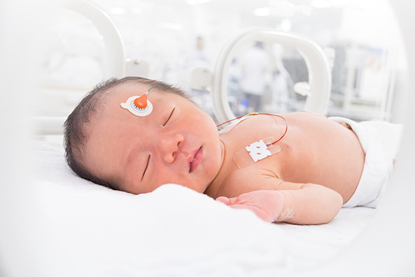
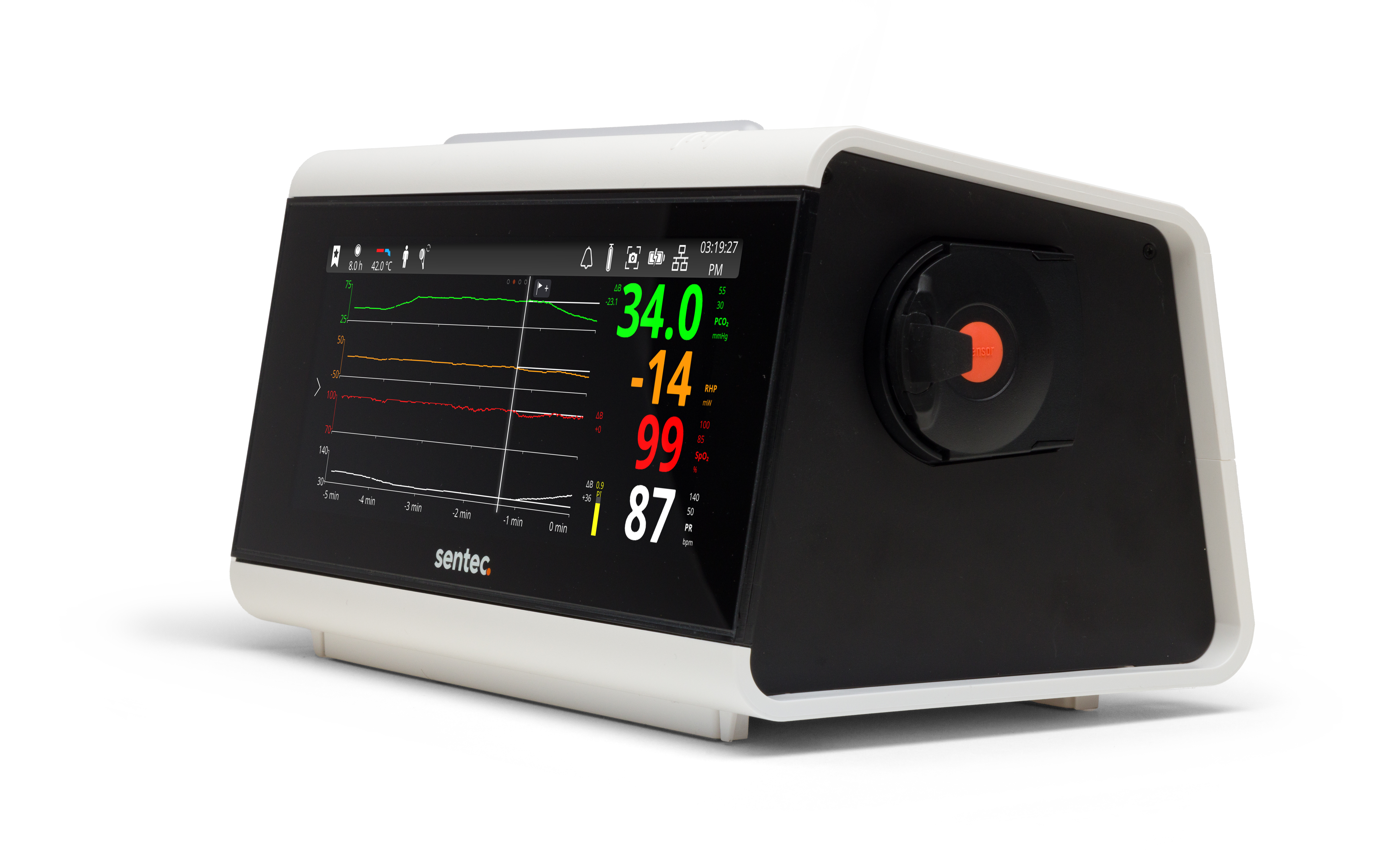
Transcutaneous CO2 (tcPCO2) monitoring is a continuous monitoring method that uses a gently heated sensor placed on the skin to estimate arterial CO2 levels. The heating element increases blood flow locally, allowing the sensor to measure CO2 that diffuses from the capillaries.
The pros:
- Noninvasive
- Provides a continuous measurement of CO2 throughout the night
- Unaffected by mouth breathing and mask leaks
- Maintains accuracy in patients with V/Q mismatch
- Compatible with NIV modalities like CPAP, NPPV, and HFNC
The limitations:
- Sensor requires regular calibration
- Poor perfusion at the monitor site can affect readings
In summary…
Transcutaneous CO2 monitoring offers sleep lab teams a valuable tool to continuously monitor ventilation throughout the night. Compared with arterial blood draws, it avoids the discomfort and single-point limitations of ABGs, and unlike end-tidal CO2 monitoring, it is less affected by factors like mouth breathing, mask leaks, or noninvasive ventilation. While it does require proper sensor placement, calibration, and adequate perfusion at the measurement site, transcutaneous monitoring can provide sleep labs with a practical option for capturing continuous ventilation trends. For many patients — especially those at risk of nocturnal hypoventilation — it may help clinicians detect changes in CO2 that could otherwise go unnoticed.
References
- Katz, S.L. Assessment of sleep-disordered breathing in pediatric neuromuscular diseases. Pediatrics. 2009.
- Nardi, J., et al. Nocturnal oximetry and transcutaneous carbon dioxide in home-ventilated neuromuscular patients. Respir Care. 2012.
- Griffon, L., et al. Home Overnight Gas Exchange for Long-Term Noninvasive Ventilation in Children. Respir Care. 2020.
- Pautrat, J., et al. Carbon dioxide levels during polygraphy in children with sleep-disordered breathing. Sleep Breath. 2015.
- Resta O., et al. Prevalence and mechanisms of diurnal hypercapnia in a sample of morbidly obese subjects with obstructive sleep apnoea. Neth J Med. 2000.
- Beaudin, A.E., et al. Contribution of hypercapnia to cognitive impairment in severe sleep-disordered breathing. J Clin Sleep Med. 2022.
- Kitajima, T., et al. Relationship Between Episodic Nocturnal Hypercapnia and History of Exacerbations in Patients with Advanced Chronic Obstructive Pulmonary Disease. Int J Chron Obstruct Pulmon Dis. 2022.
- Brillante, R., et al. An overnight increase in CO2 predicts mortality in sleep disordered breathing. Respirology. 2012.