

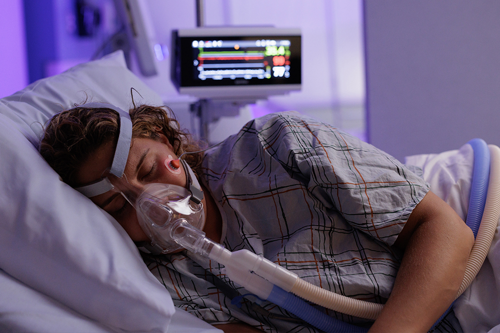
In the NICU, many infants rely on respiratory support to maintain adequate oxygenation and ventilation as their lungs grow and mature. However, that same support can also contribute to lung injury if it’s not carefully managed. The goal is to find that “just-right” level of ventilation — enough support to ensure effective gas exchange, delivered as gently as possible to protect fragile lungs. Carbon dioxide (CO2) is a key indicator of how well an infant is ventilating, making accurate CO2 monitoring valuable for guiding respiratory support and maintaining this delicate balance.
Understanding how CO2 monitoring can help fine tune respiratory support strategies, as well as the risk of abnormal levels, is important for care teams as they navigate the complexities of NICU ventilation and work to achieve the best outcomes for the smallest patients.
The role of CO2 in neonates
CO2 plays a key role in several critical physiological processes, including:
- Regulating cerebral blood flow
- Maintaining blood pH
- Triggering respiratory drive (the signal that tells the body when and how much to breathe)
- Facilitating oxygen delivery to tissues
Protecting the developing brain remains one of the highest priorities in neonatal intensive care, making the relationship between CO2 and cerebral blood flow especially important. In healthy, full-term infants, the brain can autoregulate its own blood flow. In preterm infants, however, autoregulation is immature,¹ and even small shifts in CO2 can cause harmful changes in cerebral blood flow and pressure, risking injury to the brain.2-5
Maintaining CO2 within safe ranges is therefore critical, as both hypocapnia (low CO2) and hypercapnia (high CO2) — as well as rapid fluctuations — have been shown to carry significant risks for neonates.
Hypercapnia: When CO2 is too high
Hypercapnia (also called hypercarbia) refers to above-normal CO2 levels. When CO2 levels are elevated, cerebral arteries and arterioles dilate, increasing cerebral blood flow and raising the risk of various types of brain injury.² At the same time, CO2 accumulation can lower an infant’s blood pH, contributing to additional complications.
Several studies have linked hypercapnia to adverse outcomes, including:
- Intraventricular hemorrhage (IVH)2,3,5
- Bronchopulmonary dysplasia (BPD) in low-weight infants6
- Necrotizing enterocolitis (NEC)7
- Retinopathy of prematurity (ROP)8
- Worse prognosis in infants with congenital diaphragmatic hernia (CDH)9
- Respiratory acidosis10
Hypocapnia: When CO2 is too low
Hypocapnia (also called hypocarbia) refers to below-normal CO2 levels. Hypocapnia reduces cerebral blood flow, which can increase the risk of neurological injury and other complications. It can also raise a neonate’s blood pH above normal, creating additional risks.
Several studies have linked hypocapnia to adverse outcomes, including:
- Cerebral palsy (CP)11
- Intraventricular hemorrhage (IVH)2
- Periventricular leukomalacia (PVL)12
- Cognitive developmental disorder13
- Respiratory alkalosis14
CO2 as a guide for respiratory support strategies
Many premature neonates require some form of respiratory support, such as noninvasive or invasive mechanical ventilation, due to their underdeveloped respiratory systems.15 The goal is to ensure adequate gas exchange (oxygenation and CO2 removal), while also minimizing ventilator induced lung injury (VILI). Because these infants are extremely fragile, maintaining that delicate balance is both challenging and critical for achieving the best possible outcomes.
To optimize ventilation, it is essential to monitor key gas exchange targets closely. CO2 is a direct reflection of how well ventilation matches metabolic demand, making it one of the most important parameters guiding ventilator management. Continuous CO2 monitoring not only shows whether settings are supporting effective gas exchange and helps clinicians avoid the risks of too-high or too-low CO2, but it also signals when support may be too strong — preventing potential lung overdistention and injury.
Although there is no universal consensus on the optimal pCO2 range for neonates, a recent review of the literature suggests that maintaining a pCO2 level of 37.5-52.5 mmHg is safe and effective in premature infants.7
For preterm neonates on noninvasive ventilatory support, a permissive hypercapnia strategy — maintaining PaCO2 levels around 40 – 55 mmHg — is also often used and recommended.16 This approach may reduce the need for prolonged mechanical ventilation and decrease the risk of ventilator-induced lung injury.
Optimal CO2 targets can vary depending on each infant’s condition and diagnosis. The chart below summarizes recommended CO2 target ranges for neonates, along with suggested interventions to help maintain target levels, or correct values that fall outside the desired range.

Methods of monitoring CO2 in the NICU
With the right approach to CO2 monitoring, care teams can optimize ventilation strategies while safeguarding both brain and lung health. Ongoing visibility can provide care teams with the insights they need to respond early to fluctuations, individualize care strategies, and prevent complications. Several methods are available to monitor CO2 in the NICU, each with unique strengths and limitations:
- Arterial Blood Gas (ABG): ABG tests measure CO2 and other blood gases directly from an arterial sample, providing highly accurate results and insight into acid–base balance. While considered the gold standard, the downsides of ABG tests include that they are invasive (increasing infection risk), provide only intermittent readings, contribute to pain and stimulation, and are a main contributor to blood loss in neonates.
- Capnography (End-tidal CO2): Capnography continuously measures CO2 in exhaled air, offering real-time trends and a waveform that can help identify lung compliance and airway resistance issues. While this technology is noninvasive, it is often infeasible with many patients in the NICU: uncuffed endotracheal tubes, added dead space, ventilation-perfusion (V/Q) mismatch, and low tidal volumes can cause inaccurate readings, and it is incompatible with noninvasive or high-frequency ventilation.
- Transcutaneous CO2 (tcPCO2) Monitoring: Transcutaneous CO2 monitoring is a continuous, noninvasive method where a sensor is placed on the neonate’s skin and gently heated. This heating enhances local perfusion, allowing CO2 from the blood to diffuse through the skin into the sensor, which then accurately estimates arterial CO2 levels. Although factors like poor tissue perfusion may affect accuracy in some patients, it is compatible with both noninvasive and high-frequency ventilation and is a good option for very low birth weight babies in the NICU.
Final thoughts
Effective CO2 management can support care teams in protecting infants during their most vulnerable days. Respiratory support in the NICU often requires a careful balancing act: providing enough support to maintain gas exchange, being gentle enough to safeguard fragile lungs, and avoiding the highs and lows of CO2 to protect the developing brain. CO2 serves as a key target in balancing these priorities, helping care teams identify the optimal window of support for each patient throughout their care journey.
References:
- Hoffman SB et al. J Perinatol, 2021.
- Fabres J et al. Pediatrics, 2007.
- Zayek M et al. Am J Perinatol, 2013.
- Altaany D et al. Am J Perinatol, 2015.
- Ambalavanan N et al. Arch Dis Child Fetal Neonatal Ed, 2015.
- Subramanian S et al. Matern Child Health J, 2011.
- Wong SK et al. Pediatr Res, 2022.
- Ali AA et al. Acta Paediatr, 2017.
- Abbas PI et al. J Pediatr Surg, 2015.
- Chapman K, Dragan KE. StatPearls, 2023.
- Collins MP et al. Pediatr Res, 2001.
- Shankaran S et al. Pediatrics, 2006.
- Friend AT et al. Exp Physiol, 2019.
- Sharma S, Hashmi MF. StatPearls, 2023.
- Dargaville PA et al. Pediatrics, 2016.
- Thome UH et al. Lancet Respir Med, 2015.
References:
- Hoffman SB et al. J Perinatol, 2021.
- Fabres J et al. Pediatrics, 2007.
- Zayek M et al. Am J Perinatol, 2013.
- Altaany D et al. Am J Perinatol, 2015.
- Ambalavanan N et al. Arch Dis Child Fetal Neonatal Ed, 2015.
- Subramanian S et al. Matern Child Health J, 2011.
- Wong SK et al. Pediatr Res, 2022.
- Ali AA et al. Acta Paediatr, 2017.
9. Abbas PI et al. J Pediatr Surg, 2015.
10.Chapman K, Dragan KE. StatPearls, 2023.
11. Collins MP et al. Pediatr Res, 2001.
12. Shankaran S et al. Pediatrics, 2006.
13. Friend AT et al. Exp Physiol, 2019.
14. Sharma S, Hashmi MF. StatPearls, 2023.
15. Dargaville PA et al. Pediatrics, 2016.
16. Thome UH et al. Lancet Respir Med, 2015.


")
")